The goal of the microscopic imaging core is to provide the equipment, support, and training needed by faculty and students to perform advanced cell biology, biochemistry, and histopathology studies with both fixed and live specimens. Users are encouraged to contact the instrument managers to discuss their equipment needs. 

Director
William, J. Hendry III, PhD

Equipment Managers
Raj Logan, PhD (confocal)
Kandatege Wimalasena, PhD (widefield)
Moriah Beck, PhD (TIRF)

Leica TCS SP5 II Confocal Microscopeconfocal microscope set up


The Leica TCS SP5 II is a confocal laser scanning microscope with capabilities in fluorescence imaging and 3D reconstruction. 
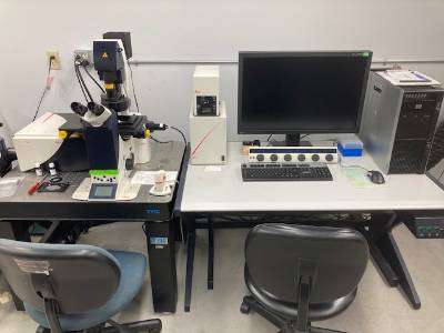
The system includes a range of
high-quality objective lenses, including oil-immersion, with different magnifications
and numerical apertures to suit various applications. It can perform
multi-dimensional imaging, including time-lapse, Z-stack, and multi-channel
imaging, enabling the acquisition of 3D datasets. It can perform spectral imaging,
allowing the separation of overlapping fluorophores by collecting full emission
spectra at each pixel. It can acquire images in multiple fluorescence channels
simultaneously, enabling colocalization studies and the observation of multiple
fluorophores within the same sample. Leica's software includes image processing
and analysis tools for data quantification and visualization. Funds to purchase
this microscope came from the Center of Innovation for Biomaterials in
Orthopaedic Research (CIBOR) at Wichita State University's National Institute for
Aviation Research.
Contact Dr. Raj Logan for training and use. 

Nikon Eclipse Ti-S

widefield microscope set up

The Nikon Eclipse Ti-S is an inverted microscope making it suitable for live cell
imaging, cell culture work, and other applications where specimens are grown
on the bottom of culture dishes or slides. The microscope includes motorized
components. These features allow for automated and precise control of the
microscope, making it suitable for multi-dimensional imaging and high-throughput
applications. It supports fluorescence microscopy with the capability to accommodate
multiple fluorescence filter sets and light sources, allowing for multi-channel
fluorescence imaging and colocalization studies. The Ti-S features high-quality
objectives and optics, ensuring excellent image quality and clarity. The Ti-S
uses Köhler illumination for optimized sample illumination. Dedicated software
for controlling and acquiring images with the Ti-S makes it easy to set up experiments
and acquire data.
Contact Dr. Kandetege Wimalasena for training and use.

Olympus TIRF

TIRF microscope set up

Total internal reflection fluorescence (TIRF) microscopy is ideal for visualizing
structures in the immediate vicinity (~100 nm) of the coverslip including cell
membrane structure and dynamics, but is also ideal for resolving single molecules
such as actin filament polymerization and cytoskeletal motility assays. The
system operates by bouncing a laser beam off of the coverslip so that photons
never hits the specimen directly; yet, the radiating evanescent wave is sufficient
to illuminate fluorescent structures nearby. The result is superior signal-to-noise
since structures outside the focal plane are never illuminated. TIRF microscopy
is also suitable for studying membrane dynamics in living cells since the laser
does not induce damage. The TIRF system consists of an Olympus IX81 with ZDC
stage control fitted with a single 488 nm laser to accommodate a wide range of
experimental demands. Phase contrast, DIC and widefield fluorescence modalities
are also available. The microscope has 10x and 20x NA 0.4 dry and 60x NA 1.45 and
63X NA 1.40 oil objectives. The EM-CCD camera is a Rolera Thunder and Metamorph
Advanced imaging software is regularly updated with ongoing support from
Olympus engineers. Funds to purchase this microscope were provided by a K-INBRE
Core Facility grant from an Institutional Development Award (IDeA) from the National
Institute of General Medical Sciences of the National Institutes of Health under grant
number P20GM103418 with matching funds from the WSU Chemistry Department,
Fairmount College of Liberal Arts and Sciences, Department of Biological Sciences,
National Center of Innovation for Biomaterials in Orthopaedic Research (CIBOR) at
Wichita State University's National Institute for Aviation Research, and M. Beck’s
start-up funds.
Contact Dr. Moriah Beck for training and use.

Nikon Eclipse E800

Widefield microscope in biology

The Nikon Eclipse E800 is an upright microscope and features an advanced optical
system designed for exceptional image clarity and brightness. A range of high-quality
objective lenses with various magnifications and numerical apertures suit different
microscopy applications. The E800 has fluorescence filter sets and fluorescence light
sources for observing fluorescently labeled samples. It supports various observation
modes, including brightfield, phase contrast, differential interference contrast (DIC),
and polarized light, in addition to fluorescence. A trinocular head enables simultaneous
observation through the eyepieces and the attachment of a camera for documentation
or imaging purposes. The microscope employs Köhler illumination, a method for optimizing
sample illumination, which helps enhance image quality.
Contact Dr. William Hendry for training and use.