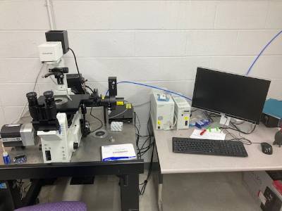
The system operates by bouncing a laser beam off of the coverslip so that photons
never hits the specimen directly; yet, the radiating evanescent wave is sufficient
to illuminate fluorescent structures nearby. The result is superior signal-to-noise
since structures outside the focal plane are never illuminated. TIRF microscopy is
also suitable for studying membrane dynamics in living cells since the laser does
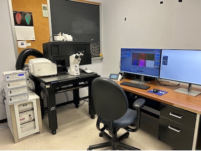
not induce damage. The TIRF system consists of an Olympus IX81 with ZDC stage control
fitted with a single 488 nm laser to accommodate a wide range of experimental demands.
Phase contrast, DIC and widefield fluorescence modalities are also available. The
microscope has 10x and 20x NA 0.4 dry and 60x NA 1.45 and 63X NA 1.40 oil objectives.
The EM-CCD camera is a Rolera Thunder and Metamorph Advanced imaging software is regularly
updated with ongoing support from Olympus engineers. Funds to purchase this microscope
were provided by a K-INBRE Core Facility grant from an Institutional Development Award
(IDeA) from the National Institute of General Medical Sciences of the National Institutes
of Health under grant number P20GM103418 with matching funds from the WSU Chemistry
Department, Fairmount College of Liberal Arts and Sciences, Department of Biological
Sciences, National Center of Innovation for Biomaterials in Orthopaedic Research (CIBOR)
at Wichita State University's National Institute for Aviation Research, and M. Beck’s
start-up funds.
Email Dr. Moriah Beck for training and use.